CLAIR Z1












Simple yet sophisticated

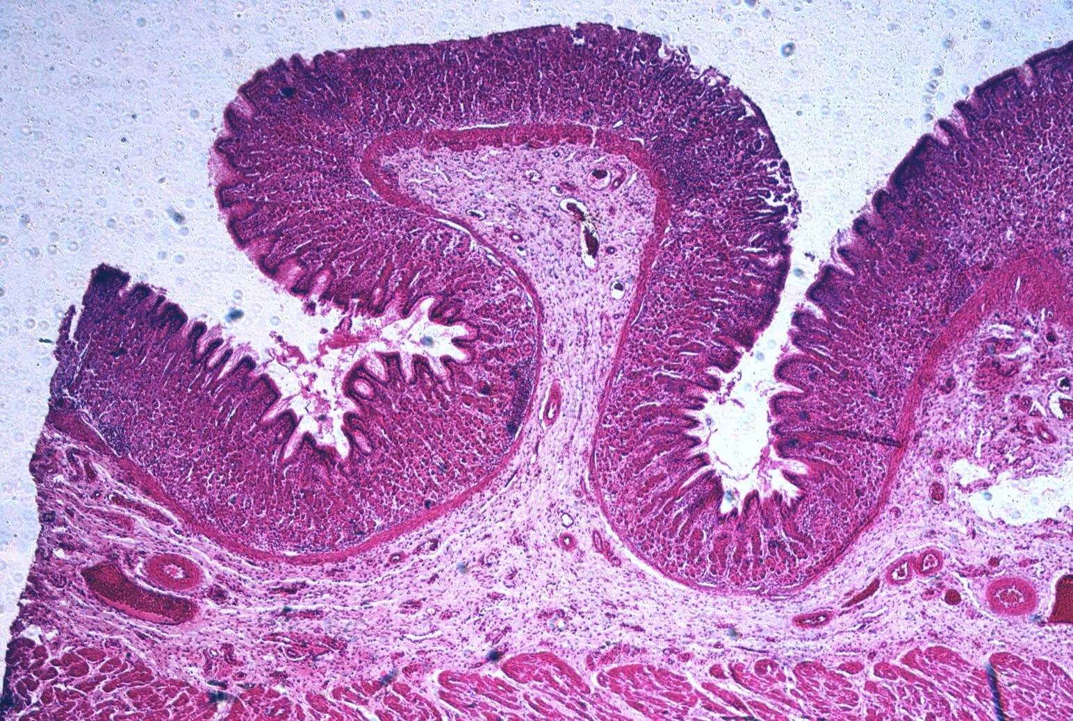
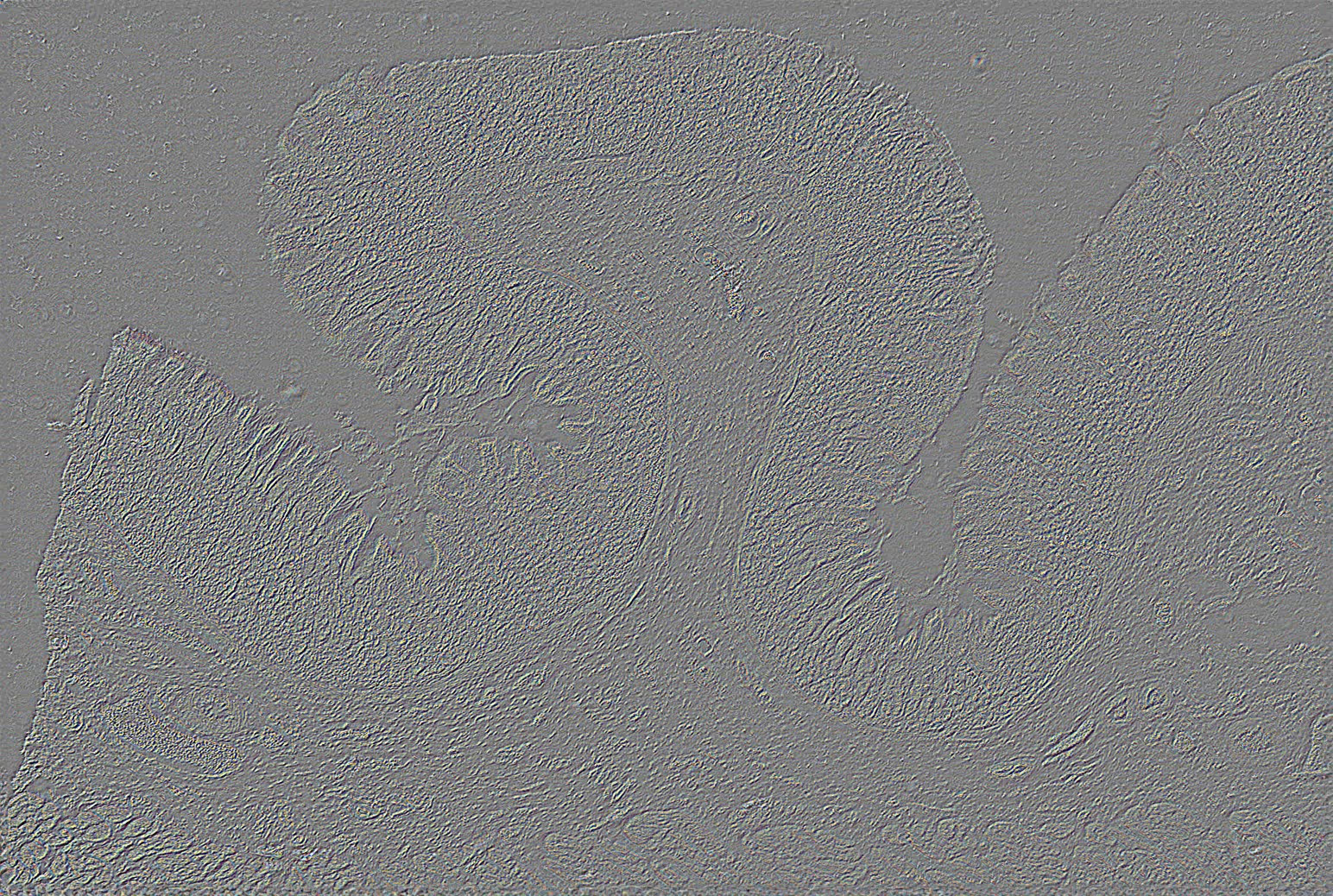
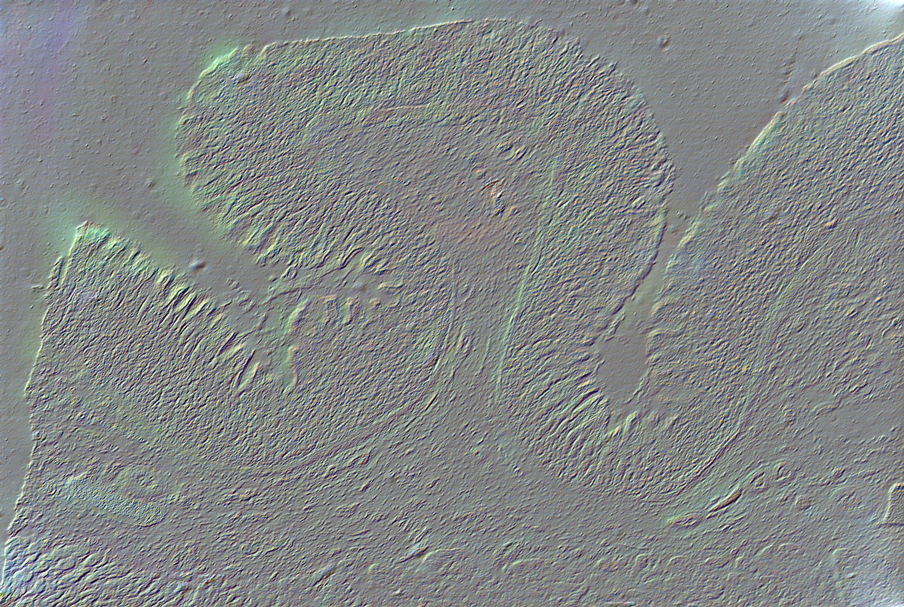
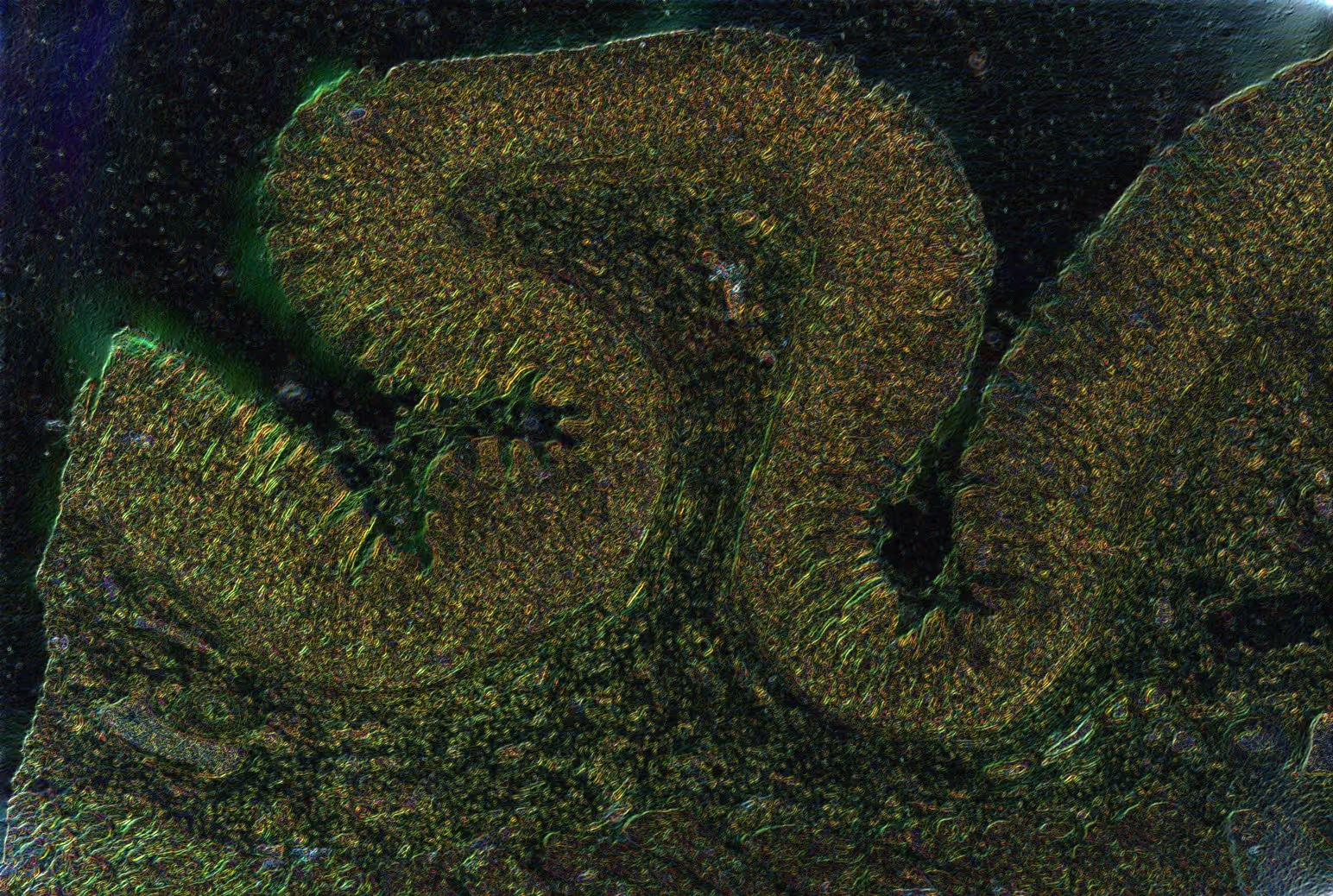

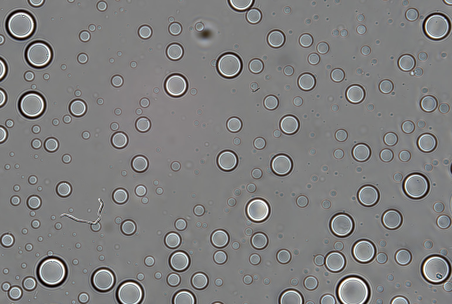


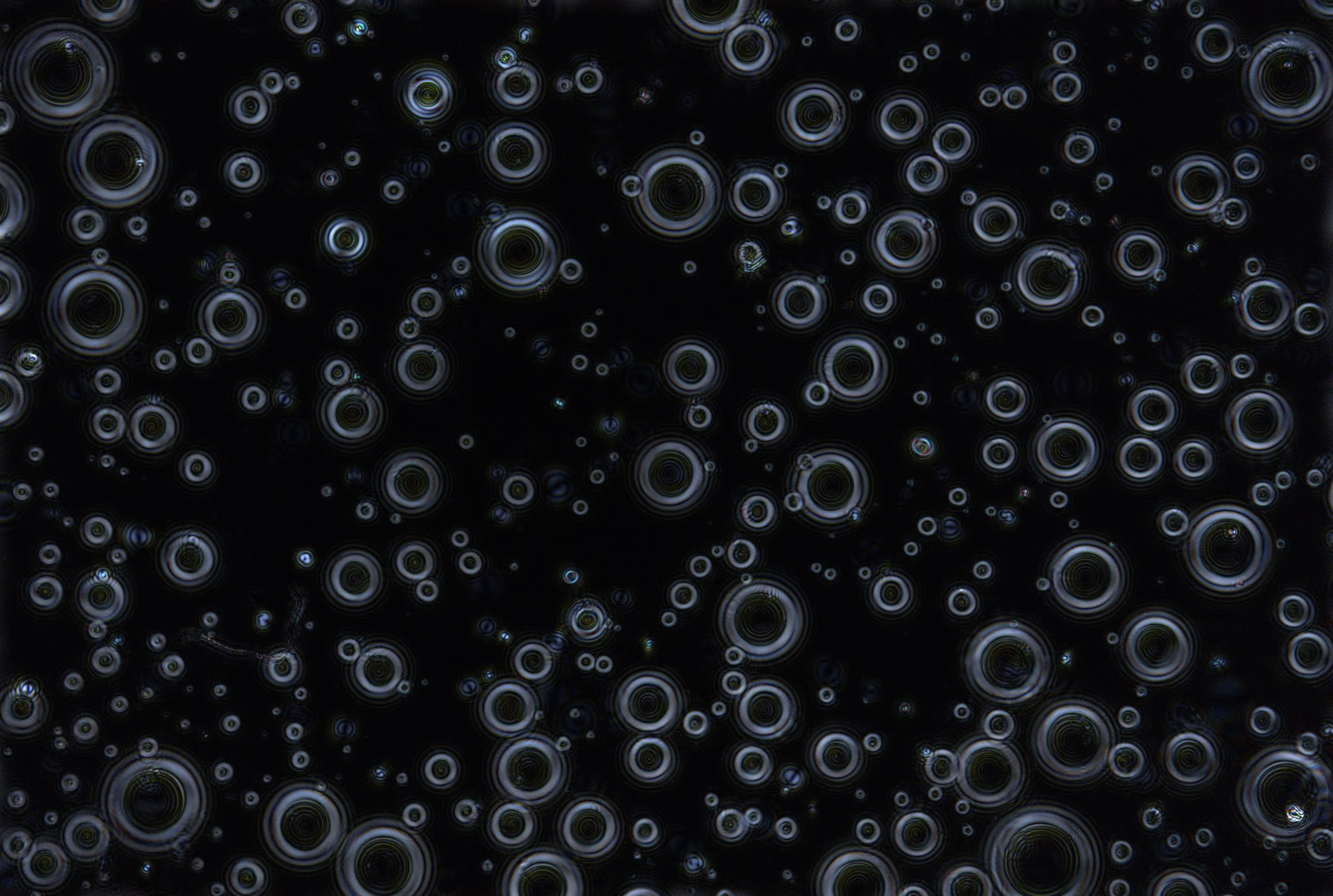

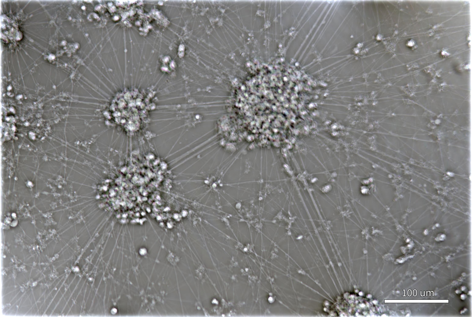
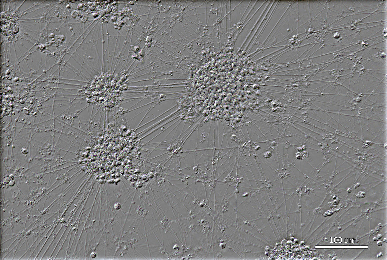
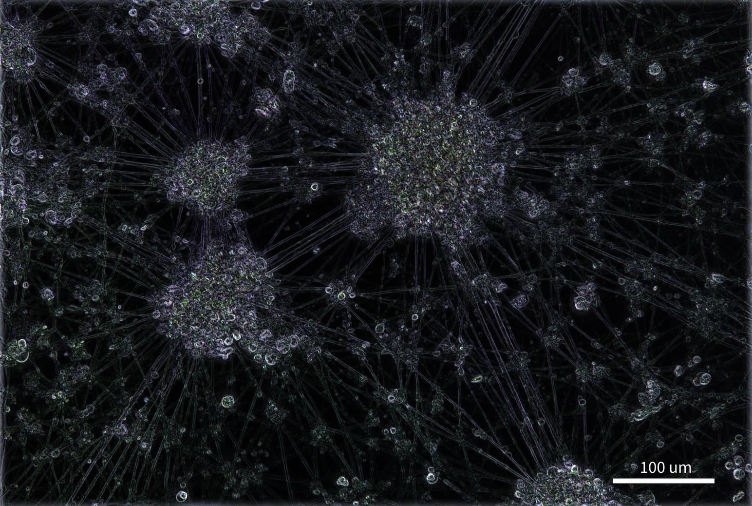


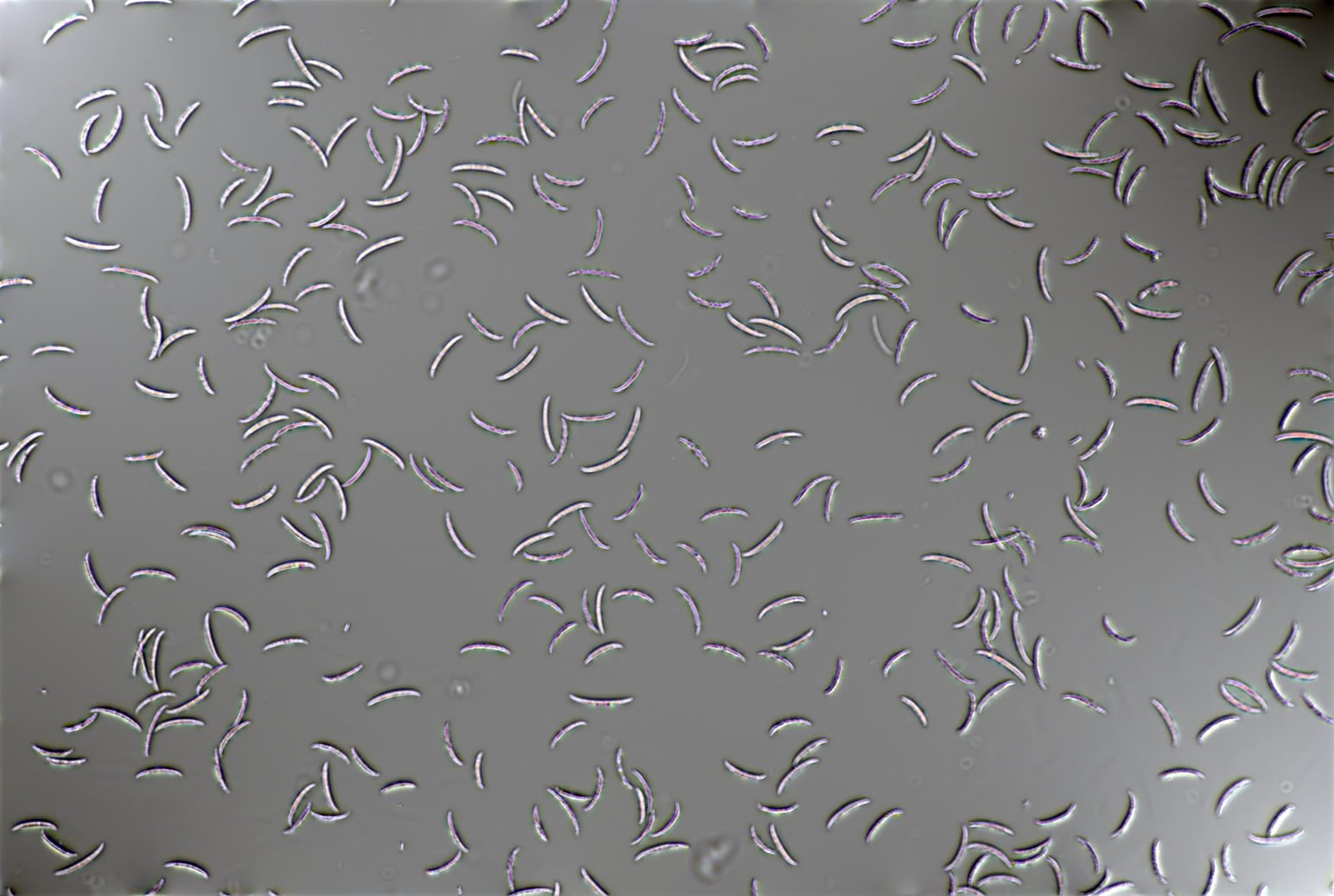
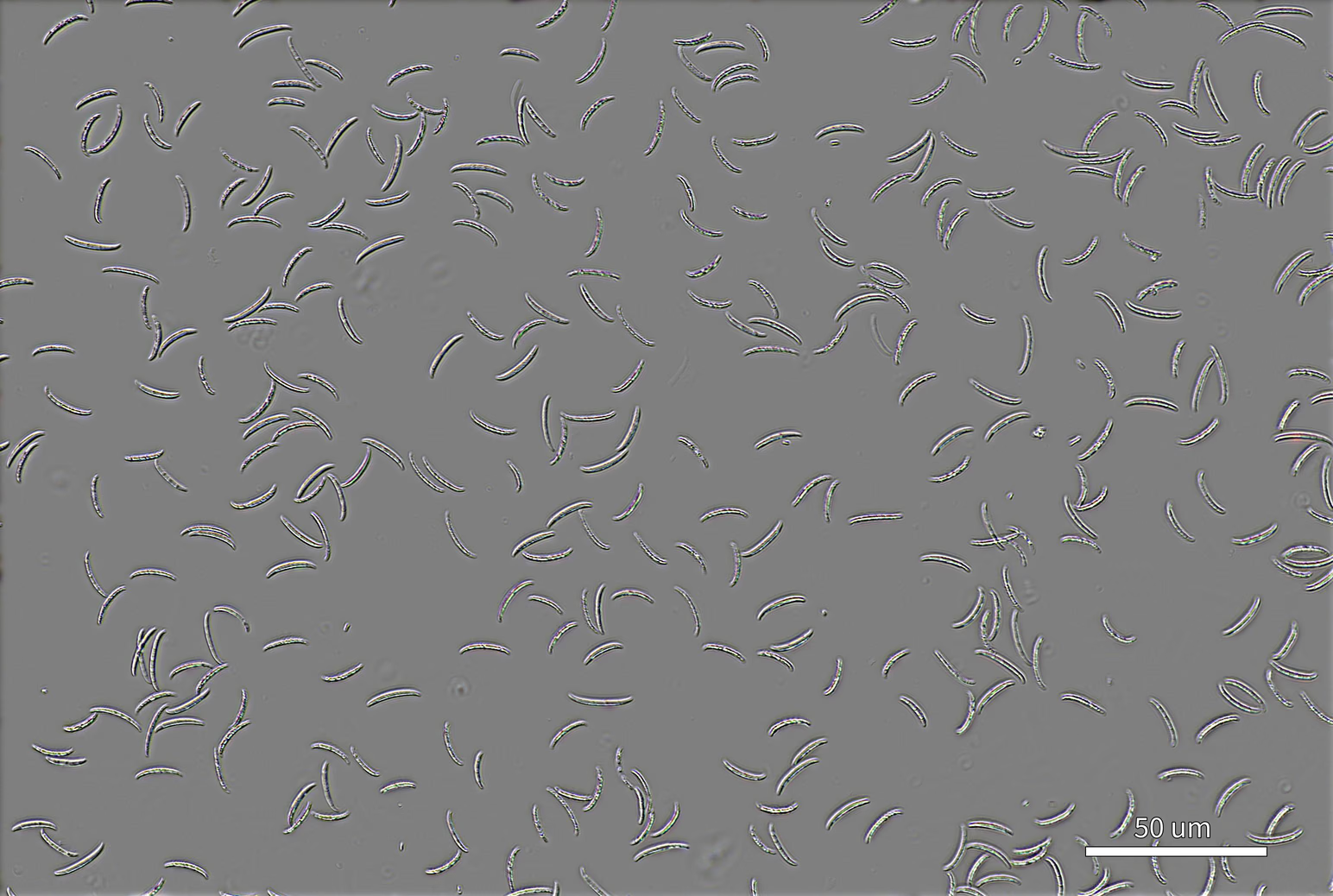


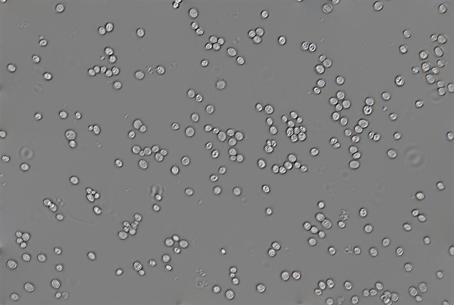
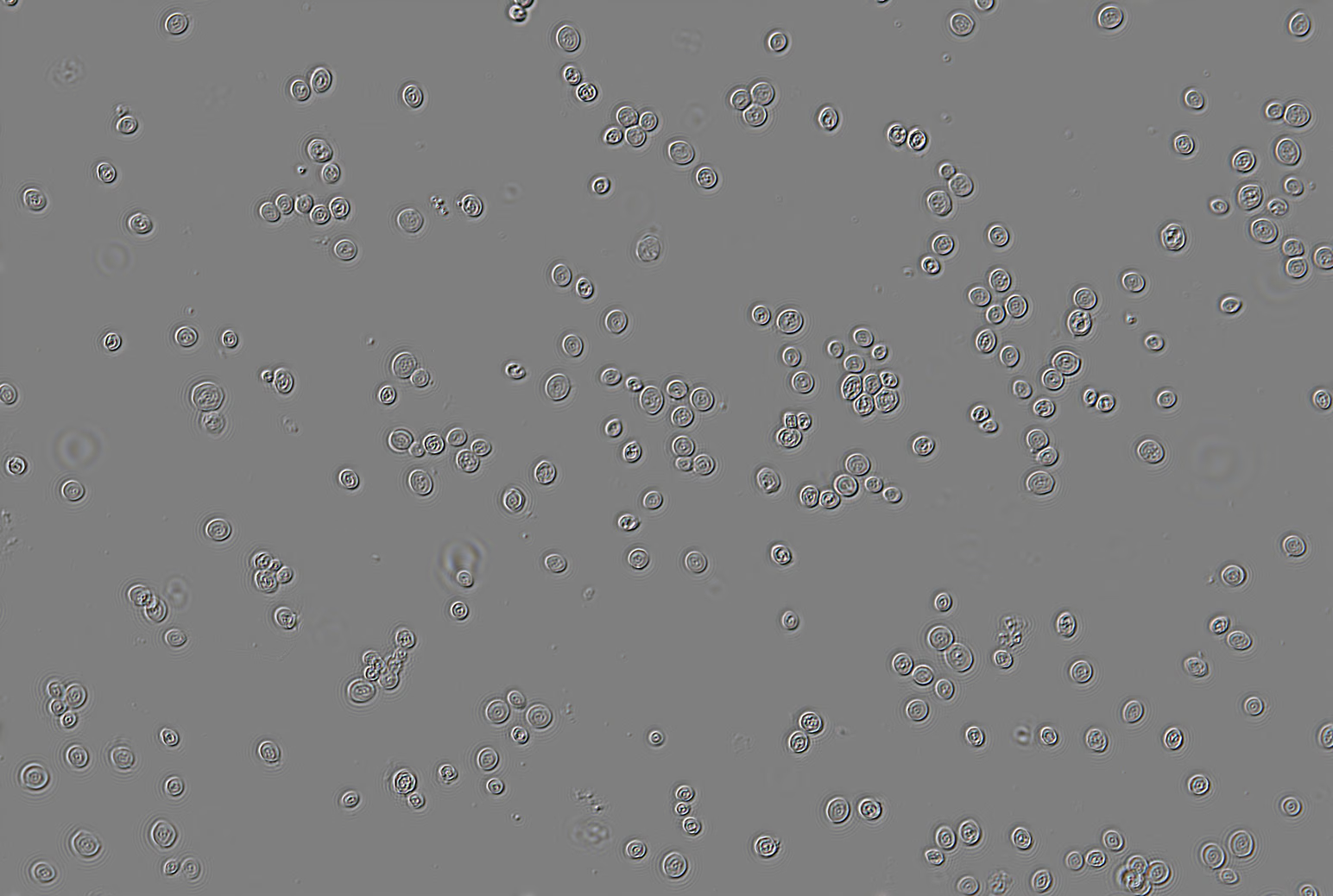

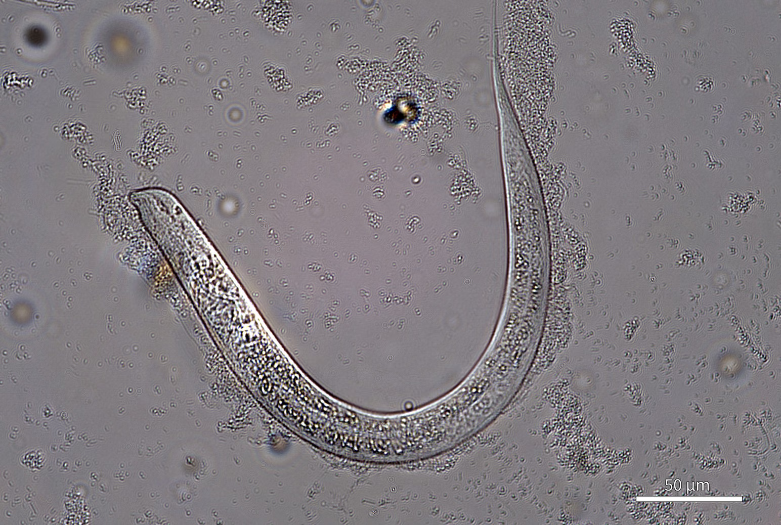
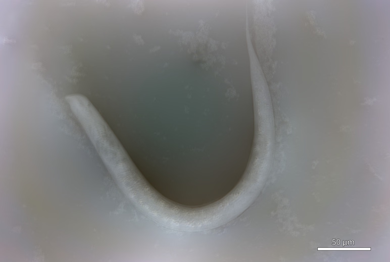
Fully featured microscopy
Designed for effortless operation, users can learn the system quickly and begin collecting reliable, high-quality data within minutes.

The Z1 is among the lightest and most space-efficient automated microscopes ever. Its small footprint means there's space for it even in crowded labs and it can fit inside a standard under-bench incubator where the Z1s integrated fan ensures consistent environmental conditions.

Get relief from repetitive microscopy tasks thanks to complete automation capabilities and compatibility with standard 85 x 125 mm multi-well microplates. Easily create automated routines for nearly every measurement imaginable, including plate-scanning, time-lapse imaging, Z/focus – stacking,. and full-slide scanning.

Open-source sample adapter designs can be downloaded, 3D printed, and modified to support custom sample formats beyond standard 85 × 125 mm assay plates. The Z1 can be operated remotely via a REST API that enables script-based control of scanning, focusing, image acquisition, and metadata management, while the built-in Windows network file share provides seamless access to data from anywhere.

Performance meets
affordability
3D characterization
Live cell imaging
Nanotechnology
… and more
Specifications

FAQ
What else do I need to get up and running?
Nothing! CLAIR Z1 can be placed on an ordinary lab bench and doesn’t require any peripherals (e.g. PCs). Some users like to connect an external monitor, but this is not required.
What labware do you support?
6 to 1536-well flat-bottomed assay plates, as well as 25x75 mm slides (including ibidi multi-well slides) and 25x25mm coverslips via our adapters. However, we encourage custom-made and 3D printedsolutions for other sample types, such as petri dishes, microfluidic flowchambers, etc.
Can the sample plates be covered?
Yes, provided the cover is optically transparent(e.g. a plate lid or sealing film). Condensation on the inside of the cover isusually not a problem.
Can I get a demonstration?
Yes, both online and in-person demos are available. → Contact Us
What are the ownership options?
We offer solutions for purchasing (from €29,000 ex-VAT) and for hiring. Please contact us for bulk-purchase pricing.
When can you deliver?
Within 10 working days, depending on delivery logistics.
Which objectives are compatible?
4x, 10x, 20x and 40x Nikon CFI Plan Fluor objectives are supported. However, please reach out if you’d like to try something different. → Contact Us
What's the resolution?
Spatial resolution is approximately 540 nm with the 40X objective lens, while phase resolution is approximately 0.01 radians. All resolution values are determined experimentally on every machine.
Do you offer service contracts and support?
Yes, we offer standard and priority service contracts and support.
Do I need a computer to use the Z1?
No, the Z1 can be fully controlled by its touchscreen alone. However, you may prefer to operate it using a phone, or a laptop, desktop, or tablet computer via its browser-based interface.
Does the Z1 have an in-built incubator or heating stage?
No, however the machine is small enough to fitinside most under-bench incubators and has been designed to withstand the environmental conditions (temperature, humidity, CO2) of most incubators.
My application is not listed; can it be supported?
Yes, probably! We work on many types of sample beyond those listed on this webpage. Please reach out to discuss your application. → Contact Us
Is it automated?
Yes, the Z1 can carry out automated scans overmultiple positions, and also z-stacks / focus-stacks as needed. Complex combinations of automated instructions can also be issued via our automation workflow or using our API.
Is photo-bleaching an issue?
Not usually. Our method uses a low illumination intensity (essentially the same as for classical brightfield microscopy). Furthermore, the illumination can be automatically switched off in-between acquisitions to further reduce light dose.
Can the Z1 do fluorescence microscopy?
No. The Z1 is a dedicated transmission light microscope. The filters, optics and mechanical components needed for fluorescence imaging in combination with quantitative phase imaging would, make the Z1 very expensive.
What's the cost of software updates?
Software updates are FREE! Machine software updates are pushed automatically over-the-air to networked instruments.